Cognitive impairments are common among patients suffering from brain tumors. Up to date, however, it is rarely assessed in clinical routine. This study aimed to evaluate pre- and postoperative neurocognitive performance in a wide range of patients suffering from gliomas representing clinical routine by using a test battery of easy-to-use and established neurocognitive tests. Patients undergoing microsurgical glioma resection between 04/2019 and 03/2021 were prospectively included. A structured test set for neurocognitive function was performed preoperatively in 33 patients and during follow-up in 14 patients. Data were converted into z-scores and combined with the corresponding cognitive domains. Thirty-three patients aged 49.2 ± 14.4 (22-81) years were included. The individual tests showed impairments preoperatively most frequently in the trail-making test B (TMT-B) in 63.6% of patients, followed by the Montreal Cognitive Assessment (MoCA) with 39.4%. Preoperatively, a clinically significant impairment was found in the domain of executive function and attention, with a mean domain score of -2.49. At follow-up, the group domain scores were impaired on the same cognitive domains as preoperatively, with executive function and attention significantly impaired (z = -2.58). Neurocognitive deficits are present in the majority of patients with glioma before surgery while still performing well in conventional scores regarding functional status. We did not observe any significant surgery-related deterioration in cognitive performance; however, this finding is compromised by a considerable number of patients lost to follow-up.
| Published in | Clinical Neurology and Neuroscience (Volume 10, Issue 1) |
| DOI | 10.11648/j.cnn.20261001.15 |
| Page(s) | 28-41 |
| Creative Commons |
This is an Open Access article, distributed under the terms of the Creative Commons Attribution 4.0 International License (http://creativecommons.org/licenses/by/4.0/), which permits unrestricted use, distribution and reproduction in any medium or format, provided the original work is properly cited. |
| Copyright |
Copyright © The Author(s), 2026. Published by Science Publishing Group |
Glioma, Cognitive Deficits, Cognitive Testing, Neurocognitive Impairments
preOP (n=33) | lost to FU (n=19) | FU (n=14) | p-value (lost to FU/ FU) | |
|---|---|---|---|---|
Gender (n (%)) | ||||
1) Male | 24 (72.7) | 13 (68.4) | 11 (78.6) | - |
2) Female | 9 (27.3) | 6 (31.6) | 3 (21.4) | |
Age | ||||
Mean ± SD (Min - Max) | 49.2±14.4 (22-81) | 53±14.7 (22-81) | 43.6±12.5 (25-72) | 0.021 |
Hemisphere (n (%)) | ||||
1) Left | 17 (51.5) | 10 (52.6) | 7 (50) | - |
2) Right | 16 (48.5) | 9 (47.4) | 7 (50) | |
Localization (n (%)) | ||||
1) Frontal | 11 (33.3) | 6 (31.6) | 5 (35.7) | - |
2) Parietal | 9 (27.3) | 6 (31.6) | 3 (21.4) | |
3) Temporal | 5 (15.15) | 2 (10.5) | 3 (21.4) | |
4) Insular | 5 (15.15) | 2 (10.5) | 3 (21.4) | |
5) Other | 3 (9.1) | 3 (15.8) | 0 (0.0) | |
WHO-Grade (n (%)) | ||||
1) 2 | 8 (24.2) | 4 (21.1) | 4 (28.6) | 0.416 |
2) 3 | 10 (30.3) | 5 (26.3) | 5 (35.7) | |
3) 4 | 15 (45.5) | 10 (52.6) | 5 (35.7) | |
Previous surgeries (n (%)) | ||||
1) Primary resection | 20 (60.6) | 10 (52.6) | 10 (71.4) | 0.310 |
2) Recurrence | 13 (39.4) | 9 (47.4) | 4 (28.6) |
preOP | preOP lost to FU | preOP with FU | FU | p-value (preOP with FU/ FU) | p-value (preOP lost to FU/ preOP with FU) | |
|---|---|---|---|---|---|---|
Number of patients (n) | KPI: 31; BI: 32; mRS: 33 | KPI: 17; BI: 16; mRS: 19 | 14 | 14 | - | - |
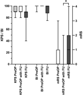
Karnofsky Performance Index (KPI) (Median (Min-Max)) | 90 (80-100) | 90 (80-100) | 90 (80-100) | 90 (40-100) | 0.055 | 0.202 |
Barthel-Index (BI) (Median (Min-Max)) | 100 (90-100) | 90 (80-100) | 100 (90-100) | 100 (75-100) | 0.125 | 0.743 |
Modified Rankin Scale (mRS) (Median (Min-Max)) | 0 (0-3) | 1 (0-4) | 0 (0-2) | 1 (0-4) | 0.012 | 0.048 |
preOP n (%) | preOP lost to FU | preOP with FU n (%) | FU n (%) | p-value (preOP with FU/FU) | p-value (preOP lost to FU/ preOP with FU) | |
|---|---|---|---|---|---|---|
Motor Function (BMRC) | 33 | 19 | 14 | 14 | ||
BMRC 5/5 | 28 (84.8) | 15 (78.9) | 13 (92.9) | 9 (64.3) | 0.125 | 0.321 |
BMRC 4/5 | 4 (12.1) | 3 (15.8) | 1 (7.1) | 4 (28.6) | ||
BMRC <4/5 | 1 (3.1) | 1 (5.3) | 0 | 1 (7.1) |
Test | preOP | Primary tumor (preOP) | Recurrent tumor (preOP) | preOP lost to FU | preOP with FU | FU | |||||||
|---|---|---|---|---|---|---|---|---|---|---|---|---|---|
n | Impaired (n (%)) | n | Impaired (n (%)) | n | Impaired (n (%)) | n | Impaired (n (%)) | n | Impaired (n (%)) | n | Impaired (n (%)) | Cut-off | |
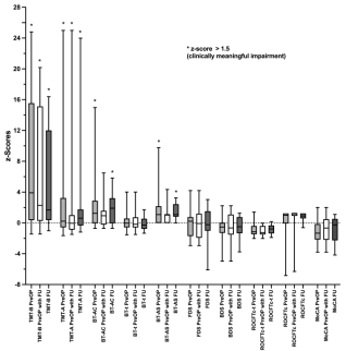
MoCA | 33 | 13 (39.4) | 20 | 9 (45.0) | 13 | 4 (30.8) | 19 | 9 (47.4) | 14 | 4 (28.6) | 14 | 6 (42.9) | * |
TMT-A | 33 | 12 (36.4) | 20 | 7 (35.0) | 13 | 5 (38.5) | 19 | 9 (47.4 | 14 | 3 (21.4) | 14 | 4 (28.6) | * |
TMT-B | 33 | 21 (63.6) | 20 | 13 (65.0) | 13 | 8 (61.5) | 19 | 13 (68.4) | 14 | 8 (57.1) | 14 | 7 (50.0) | * |
BT | 33 | 20 | 13 | 19 | 14 | 14 | |||||||
BT-AC | 12 (36.4) | 4 (20.0) | 8 (61.5) | 9 (47.4) | 3 (21.0) | 8 (57.1) | * | ||||||
BT-AS | 6 (18.2) | 4 (20.0) | 2 (15.4) | 5 (26.3) | 1 (7.1) | 3 (21.4) | ≥3 | ||||||
BT-t | 2 (6.1) | 1 (5.0) | 1 (7.7) | 1 (5.3) | 1 (7.1) | 2 (14.3) | * | ||||||
LB | |||||||||||||
Score | 33 | 5 (15.2) | 20 | 4 (20.0) | 13 | 1 (7.7) | 19 | 3 (15.8) | 14 | 2 (14.3) | 14 | 4 (28.6) | ≤14 ** |
FBDS | 33 | 20 | 13 | 19 | 14 | 14 | |||||||
FDS | 9 (27.3) | 5 (25.0) | 4 (30.8) | 4 (21.1) | 5 (35.7) | 3 (21.4) | * | ||||||
BDS | 5 (15.2) | 4 (20.0) | 1 (7.7) | 2 (10.5) | 3 (21.4) | 2 (14.3) | * | ||||||
ROCFTc | 31 | 20 | 13 | 17 | 14 | 14 | |||||||
Score | 4 (12.1) | 4 (20.0) | 0 | 3 (17.6) | 1 (7.1) | 0 | * | ||||||
Time | 0 (0.0) | 0 | 0 | 0 | 0 | 0 | * | ||||||
QAB | 12 | 1 (8.33) | 5 | 1 (20.0) | 7 | 0 | 10 | 1 (10.0) | 2 | 0 | 3 | 2 (66.7) | <8.9 |
Domains and tests | n (preOP) | Z-scores mean (SD) preOP | Group domain scores preOP | n (preOP lost to FU) | Z-scores mean (SD) preOP lost to FU | Group domain scores preOP lost to FU | n (preOP with FU) | Z-scores mean (SD) preOP with FU | Group domain scores preOP with FU | n (FU) | Z-scores mean (SD) FU | Group domain scores FU |
|---|---|---|---|---|---|---|---|---|---|---|---|---|
Executive and attention | ||||||||||||
TMT-B | 33 | 7.30 (7.77) | -3.45 | 19 | 8.53 (7.82) | -4.16 | 14 | 5.63 (7.65) | -2.49 | 14 | 5.24 (6.57) | -2.58 |
BDS | 33 | -0.73 (1.61) | 19 | -0.80 (1.19) | 14 | -0.64 (2.11) | 14 | -0.49 (1.38) | ||||
BT-AC | 33 | 2.32 (3.61) | 19 | 3.15 (4.27) | 14 | 1.21 (2.10) | 14 | 2.0 (1.99) | ||||
Memory | ||||||||||||
BDS | 33 | -0.73 (1.61) | -0.40 | 19 | -0.80 (1.19) | -0.43 | 14 | -0.64 (2.11) | -0.36 | 14 | -0.49 (1.38) | -0.35 |
FDS | 33 | -0.06 (1.76) | 19 | -0.06 (1.51) | 14 | -0.07 (2.11) | 14 | -0.20 (2.16) | ||||
Visuospatial functioning | ||||||||||||
BT-AC BT-AS ROCFTc | 33 | 2.32 (3.61) | -1.24 | 19 | 3.15 (4.27) | -1.75 | 14 | 1.21 (2.10) | -0.56 | 14 | 2.0 (1.99) | -0.94 |
33 | 1.54 (1.88) | 19 | 1.94 (2.22) | 14 | 1.0 (1.16) | 14 | 1.55 (1.18) | |||||
31 | 0.15 (2.1) | 17 | -0.16 (2.18) | 14 | 0.52 (2.01) | 14 | 0.74 (0.6) | |||||
Processing speed | ||||||||||||
TMT-A | 33 | 2.46 (5.66) | -0.57 | 19 | 2.65 (4.57) | -0.68 | 14 | 2.22 (7.05) | -0.44 | 14 | 2.41 (6.43) | -0.51 |
ROCFTc-t | 31 | -0.87 (0.91) | 17 | -0.70 (1.08) | 14 | -1.06 (0.64) | 14 | -0.83 (0.65) | ||||
BT-t | 33 | 0.12 (1.24) | 19 | 0.10 (1.18) | 14 | 0.16 (1.36) | 14 | -0.06 (0.97) | ||||
General cognition | ||||||||||||
MoCA | 33 | -1.04 (1.43) | - | 19 | -1.20 (1.36) | - | 14 | -0.83 (1.54) | - | 14 | -0.92 (1.71) | - |
AT | Adjuvant Therapy |
BDS | Backwards Digit Span |
BI | Barthel Index |
BMRC | British Medical Research Council |
BT-AC | Bells Test's Accuracy Score |
BT-AS | Bells Test's Asymmetry Score |
BT-t | Bells Test Task Completion Time |
FDS | Forwards Digit Span |
FU | Follow-up |
HGG | High-grade Glioma |
KPS | Karnofsky Performance Scale |
LB | Line Bisection |
LGG | Low-grade Glioma |
Lost to FU | Lost to Follow-up Cohort |
MoCa | Montreal Cognitive Assessment |
MRI | Magnetic Resonance Imaging |
mRS | Modified Rankin Scale |
nonAT | Non-adjuvant Therapy |
preOP with FU | Preoperative Test Result of the Follow-up Cohort |
QAB | Quick Aphasia Battery |
ROCFTc | Copying task of the Rey-Osterrieth-Complex-Figure Test |
ROCFTc-t | Task Completion Time of the ROCFTc-t |
TMT-A | Trail Making Test Part A |
TMT-B | Trail Making Test Part B |
WHO CNS° | Tumor Grades According to the World Health Organisation Classification of Tumors of the Central Nervous System |
| [1] | Gehring K, Taphoorn MJ, Sitskoorn MM, Aaronson NK (2015) Predictors of subjective versus objective cognitive functioning in patients with stable grades II and III glioma. Neurooncol Pract 2: 20-31. |
| [2] | Pertz M, Kowalski T, Jetschke K, Schmieder K, Schlegel U, Miller D (2022) Pre- and postoperative self-reported and objectively assessed neurocognitive functioning in lower grade glioma patients. J Clin Neurosci 106: 185-193. |
| [3] | Fuster JM (2006) The cognit: a network model of cortical representation. Int J Psychophysiol 60: 125-132. |
| [4] | Kanai R, Rees G (2011) The structural basis of inter-individual differences in human behaviour and cognition. Nat Rev Neurosci 12: 231-242. |
| [5] | Humphreys GF, Lambon Ralph MA (2015) Fusion and Fission of Cognitive Functions in the Human Parietal Cortex. Cereb Cortex 25: 3547-3560. |
| [6] | Habets EJ, Kloet A, Walchenbach R, Vecht CJ, Klein M, Taphoorn MJ (2014) Tumour and surgery effects on cognitive functioning in high-grade glioma patients. Acta Neurochir (Wien) 156: 1451-1459. |
| [7] | Tucha O, Smely C, Preier M, Lange KW (2000) Cognitive deficits before treatment among patients with brain tumors. Neurosurgery 47: 324-333; discussion 333-324. |
| [8] | Wilson SM, Eriksson DK, Schneck SM, Lucanie JM (2018) A quick aphasia battery for efficient, reliable, and multidimensional assessment of language function. PLoS One 13: e0192773. |
| [9] | Nasreddine ZS, Phillips NA, Bédirian V, Charbonneau S, Whitehead V, Collin I, Cummings JL, Chertkow H (2005) The Montreal Cognitive Assessment, MoCA: a brief screening tool for mild cognitive impairment. J Am Geriatr Soc 53: 695-699. |
| [10] | Reitan RM (1955) The relation of the trail making test to organic brain damage. J Consult Psychol 19: 393-394. |
| [11] | Gauthier L, Dehaut F, Joanette Y (1989) The Bells Test: A quantitative and qualitative test for visual neglect. International Journal of Clinical Neuropsychology 11: 49-54. |
| [12] | Milner AD, Harvey M, Roberts RC, Forster SV (1993) Line bisection errors in visual neglect: misguided action or size distortion? Neuropsychologia 31: 39-49. |
| [13] | Schenkenberg T, Bradford DC, Ajax ET (1980) Line bisection and unilateral visual neglect in patients with neurologic impairment. Neurology 30: 509-517. |
| [14] | Humpstone HJ (1919) Memory Span Tests. Psychol Clin 12: 196-200. |
| [15] | Osterrieth PA (1944) Le test de copie d'une figure complexe; contribution à l'étude de la perception et de la mémoire. [Test of copying a complex figure; contribution to the study of perception and memory.]. Archives de Psychologie 30: 206-356. |
| [16] | Thomann AE, Goettel N, Monsch RJ, Berres M, Jahn T, Steiner LA, Monsch AU (2018) The Montreal Cognitive Assessment: Normative Data from a German-Speaking Cohort and Comparison with International Normative Samples. J Alzheimers Dis 64: 643-655. |
| [17] | Monaco M, Costa A, Caltagirone C, Carlesimo GA (2013) Forward and backward span for verbal and visuo-spatial data: standardization and normative data from an Italian adult population. Neurol Sci 34: 749-754. |
| [18] | Tombaugh TN (2004) Trail Making Test A and B: normative data stratified by age and education. Arch Clin Neuropsychol 19: 203-214. |
| [19] | Shin MS, Park SY, Park SR, Seol SH, Kwon JS (2006) Clinical and empirical applications of the Rey-Osterrieth Complex Figure Test. Nat Protoc 1: 892-899. |
| [20] | Youn YC, Pyun JM, Ryu N, Baek MJ, Jang JW, Park YH, Ahn SW, Shin HW, Park KY, Kim SY (2021) Use of the Clock Drawing Test and the Rey-Osterrieth Complex Figure Test-copy with convolutional neural networks to predict cognitive impairment. Alzheimers Res Ther 13: 85. |
| [21] | Canham RS, Stephen & Tyrrell, Andy (2000) Automated Scoring of a Neuropsychological Test: The Rey Osterrieth Complex Figure. |
| [22] | Tremblay MP, Potvin O, Callahan BL, Belleville S, Gagnon JF, Caza N, Ferland G, Hudon C, Macoir J (2015) Normative data for the Rey-Osterrieth and the Taylor complex figure tests in Quebec-French people. Arch Clin Neuropsychol 30: 78-87. |
| [23] | Mancuso M, Damora A, Abbruzzese L, Navarrete E, Basagni B, Galardi G, Caputo M, Bartalini B, Bartolo M, Zucchella C, Carboncini MC, Dei S, Zoccolotti P, Antonucci G, De Tanti A (2018) A New Standardization of the Bells Test: An Italian Multi-Center Normative Study. Front Psychol 9: 2745. |
| [24] | Louis DN, Perry A, Wesseling P, Brat DJ, Cree IA, Figarella-Branger D, Hawkins C, Ng HK, Pfister SM, Reifenberger G, Soffietti R, von Deimling A, Ellison DW (2021) The 2021 WHO Classification of Tumors of the Central Nervous System: a summary. Neuro Oncol 23: 1231-1251. |
| [25] | Liouta E, Koutsarnakis C, Komaitis S, Kalyvas AV, Drosos E, Garcia-Gomez JM, Juan-Albarracin J, Katsaros V, Stavrinou L, Stranjalis G (2023) Preoperative neurocognitive function as an independent survival prognostic marker in primary glioblastoma. Neurooncol Pract 10: 527-535. |
| [26] | Forster MT, Behrens M, Lortz I, Conradi N, Senft C, Voss M, Rauch M, Seifert V (2020) Benefits of glioma resection in the corpus callosum. Sci Rep 10: 16630. |
| [27] | Talacchi A, Santini B, Savazzi S, Gerosa M (2011) Cognitive effects of tumour and surgical treatment in glioma patients. J Neurooncol 103: 541-549. |
| [28] | Noll KR, Sullaway C, Ziu M, Weinberg JS, Wefel JS (2015) Relationships between tumor grade and neurocognitive functioning in patients with glioma of the left temporal lobe prior to surgical resection. Neuro Oncol 17: 580-587. |
| [29] | Bosma I, Vos MJ, Heimans JJ, Taphoorn MJ, Aaronson NK, Postma TJ, van der Ploeg HM, Muller M, Vandertop WP, Slotman BJ, Klein M (2007) The course of neurocognitive functioning in high-grade glioma patients. Neuro Oncol 9: 53-62. |
| [30] | Meyers CA, Hess KR (2003) Multifaceted end points in brain tumor clinical trials: cognitive deterioration precedes MRI progression. Neuro Oncol 5: 89-95. |
| [31] | Klein M, Duffau H, De Witt Hamer PC (2012) Cognition and resective surgery for diffuse infiltrative glioma: an overview. J Neurooncol 108: 309-318. |
| [32] | Kaleita TA, Wellisch DK, Cloughesy TF, Ford JM, Freeman D, Belin TR, Goldman J (2004) Prediction of neurocognitive outcome in adult brain tumor patients. J Neurooncol 67: 245-253. |
| [33] | Satoer D, Visch-Brink E, Dirven C, Vincent A (2016) Glioma surgery in eloquent areas: can we preserve cognition? Acta Neurochir (Wien) 158: 35-50. |
| [34] | Satoer D, Visch-Brink E, Smits M, Kloet A, Looman C, Dirven C, Vincent A (2014) Long-term evaluation of cognition after glioma surgery in eloquent areas. J Neurooncol 116: 153-160. |
| [35] | Greene-Schloesser D, Robbins ME (2012) Radiation-induced cognitive impairment--from bench to bedside. Neuro Oncol 14 Suppl 4: iv37-44. |
| [36] | Greene-Schloesser D, Moore E, Robbins ME (2013) Molecular pathways: radiation-induced cognitive impairment. Clin Cancer Res 19: 2294-2300. |
| [37] | Connor M, Karunamuni R, McDonald C, Seibert T, White N, Moiseenko V, Bartsch H, Farid N, Kuperman J, Krishnan A, Dale A, Hattangadi-Gluth JA (2017) Regional susceptibility to dose-dependent white matter damage after brain radiotherapy. Radiother Oncol 123: 209-217. |
| [38] | Kirkman MA, Hunn BHM, Thomas MSC, Tolmie AK (2022) Influences on cognitive outcomes in adult patients with gliomas: A systematic review. Front Oncol 12: 943600. |
| [39] | Laack NN, Brown PD, Ivnik RJ, Furth AF, Ballman KV, Hammack JE, Arusell RM, Shaw EG, Buckner JC, North Central Cancer Treatment G (2005) Cognitive function after radiotherapy for supratentorial low-grade glioma: a North Central Cancer Treatment Group prospective study. Int J Radiat Oncol Biol Phys 63: 1175-1183. |
| [40] | Nakada M, Nakajima R, Okita H, Nakade Y, Yuno T, Tanaka S, Kinoshita M (2021) Awake surgery for right frontal lobe glioma can preserve visuospatial cognition and spatial working memory. J Neurooncol 151: 221-230. |
| [41] | Motomura K, Chalise L, Ohka F, Aoki K, Tanahashi K, Hirano M, Nishikawa T, Wakabayashi T, Natsume A (2018) Supratotal Resection of Diffuse Frontal Lower Grade Gliomas with Awake Brain Mapping, Preserving Motor, Language, and Neurocognitive Functions. World Neurosurg 119: 30-39. |
| [42] | Duffau H (2010) Awake surgery for nonlanguage mapping. Neurosurgery 66: 523-528; discussion 528-529. |
| [43] | Sarubbo S, Tate M, De Benedictis A, Merler S, Moritz-Gasser S, Herbet G, Duffau H (2020) Mapping critical cortical hubs and white matter pathways by direct electrical stimulation: an original functional atlas of the human brain. Neuroimage 205: 116237. |
| [44] | Herbet G (2021) Should Complex Cognitive Functions Be Mapped With Direct Electrostimulation in Wide-Awake Surgery? A Network Perspective. Front Neurol 12: 635439. |
APA Style
Schwendner, M., Markwardt, O., Meyer, B., Krieg, S. M., Ille, S. (2026). The Impact of Glioma Growth and Resection on Neurocognitive Impairment – a Prospective Observational Study. Clinical Neurology and Neuroscience, 10(1), 28-41. https://doi.org/10.11648/j.cnn.20261001.15
ACS Style
Schwendner, M.; Markwardt, O.; Meyer, B.; Krieg, S. M.; Ille, S. The Impact of Glioma Growth and Resection on Neurocognitive Impairment – a Prospective Observational Study. Clin. Neurol. Neurosci. 2026, 10(1), 28-41. doi: 10.11648/j.cnn.20261001.15
@article{10.11648/j.cnn.20261001.15,
  author = {Maximilian Schwendner and Odilia Markwardt and Bernhard Meyer and Sandro Manuel Krieg and Sebastian Ille},
  title = {The Impact of Glioma Growth and Resection on Neurocognitive Impairment – a Prospective Observational Study},
  journal = {Clinical Neurology and Neuroscience},
  volume = {10},
  number = {1},
  pages = {28-41},
  doi = {10.11648/j.cnn.20261001.15},
  url = {https://doi.org/10.11648/j.cnn.20261001.15},
  eprint = {https://article.sciencepublishinggroup.com/pdf/10.11648.j.cnn.20261001.15},
  abstract = {Cognitive impairments are common among patients suffering from brain tumors. Up to date, however, it is rarely assessed in clinical routine. This study aimed to evaluate pre- and postoperative neurocognitive performance in a wide range of patients suffering from gliomas representing clinical routine by using a test battery of easy-to-use and established neurocognitive tests. Patients undergoing microsurgical glioma resection between 04/2019 and 03/2021 were prospectively included. A structured test set for neurocognitive function was performed preoperatively in 33 patients and during follow-up in 14 patients. Data were converted into z-scores and combined with the corresponding cognitive domains. Thirty-three patients aged 49.2 ± 14.4 (22-81) years were included. The individual tests showed impairments preoperatively most frequently in the trail-making test B (TMT-B) in 63.6% of patients, followed by the Montreal Cognitive Assessment (MoCA) with 39.4%. Preoperatively, a clinically significant impairment was found in the domain of executive function and attention, with a mean domain score of -2.49. At follow-up, the group domain scores were impaired on the same cognitive domains as preoperatively, with executive function and attention significantly impaired (z = -2.58). Neurocognitive deficits are present in the majority of patients with glioma before surgery while still performing well in conventional scores regarding functional status. We did not observe any significant surgery-related deterioration in cognitive performance; however, this finding is compromised by a considerable number of patients lost to follow-up.},
 year = {2026}
}
TY  - JOUR
T1  - The Impact of Glioma Growth and Resection on Neurocognitive Impairment – a Prospective Observational Study
AU  - Maximilian Schwendner
AU  - Odilia Markwardt
AU  - Bernhard Meyer
AU  - Sandro Manuel Krieg
AU  - Sebastian Ille
Y1  - 2026/03/12
PY  - 2026
N1  - https://doi.org/10.11648/j.cnn.20261001.15
DO  - 10.11648/j.cnn.20261001.15
T2  - Clinical Neurology and Neuroscience
JF  - Clinical Neurology and Neuroscience
JO  - Clinical Neurology and Neuroscience
SP  - 28
EP  - 41
PB  - Science Publishing Group
SN  - 2578-8930
UR  - https://doi.org/10.11648/j.cnn.20261001.15
AB  - Cognitive impairments are common among patients suffering from brain tumors. Up to date, however, it is rarely assessed in clinical routine. This study aimed to evaluate pre- and postoperative neurocognitive performance in a wide range of patients suffering from gliomas representing clinical routine by using a test battery of easy-to-use and established neurocognitive tests. Patients undergoing microsurgical glioma resection between 04/2019 and 03/2021 were prospectively included. A structured test set for neurocognitive function was performed preoperatively in 33 patients and during follow-up in 14 patients. Data were converted into z-scores and combined with the corresponding cognitive domains. Thirty-three patients aged 49.2 ± 14.4 (22-81) years were included. The individual tests showed impairments preoperatively most frequently in the trail-making test B (TMT-B) in 63.6% of patients, followed by the Montreal Cognitive Assessment (MoCA) with 39.4%. Preoperatively, a clinically significant impairment was found in the domain of executive function and attention, with a mean domain score of -2.49. At follow-up, the group domain scores were impaired on the same cognitive domains as preoperatively, with executive function and attention significantly impaired (z = -2.58). Neurocognitive deficits are present in the majority of patients with glioma before surgery while still performing well in conventional scores regarding functional status. We did not observe any significant surgery-related deterioration in cognitive performance; however, this finding is compromised by a considerable number of patients lost to follow-up.
VL  - 10
IS  - 1
ER  -
Department of Neurosurgery Heidelberg University Hospital, Ruprecht-Karls-University Heidelberg, Heidelberg, Germany
Department of Neurosurgery Klinikum Rechts der Isar, Technische Universität München, Munich, Germany;Department of Neurology, University of Cagliari, Cagliari, Italy
Department of Neurosurgery Klinikum Rechts der Isar, Technische Universität München, Munich, Germany
Department of Neurosurgery Heidelberg University Hospital, Ruprecht-Karls-University Heidelberg, Heidelberg, Germany
Department of Neurosurgery Heidelberg University Hospital, Ruprecht-Karls-University Heidelberg, Heidelberg, Germany